• Home
• Research
• Projects
• Patents
• Press
• Demos
• Code
• Teaching
• Talks
• People
• Contact
• CV
• Music
Demos
Optics
Mouse Vision vs. Primate Vision
Astigmatism
Chromatic Aberration
Mouse Optics vs. Primate Optics
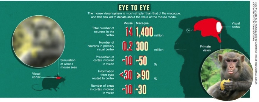
The images in this figure were created to reflect what a mouse and primate would see at a viewing distance of 2.4 meters. To simulate what each species would see, size the photographs so that your index finger at arm's length just covers both eyes of the monkey (apprx. 1 visual deg). To create the image on the left, we simulated the optics of the mouse eye to obtain the mouse's retinal image. We then sampled that image with a mosaic of cone photoreceptors having the spacing of the those in the mouse retina. We then removed the contribution of the long wavelength (L cones), because the mouse (like most mammals) has only middle (M) and short (S) wavelength cones. We did this by converting the image to the Lab color space and then removing the a (red‐green) channel. Rhesus macaque monkeys average about 21 inches (53 cm) in length, and the face length is about 1/5 of that (10.5 cm). In the image, the face is about 2.5 deg in visual angle. Thus, the viewing distance corresponds to about 2.4 meters.
Astigmatism
Approximately 15% of American adults have astigmatism that is strong enough to require refractive correction. Astigmatism is an optical defect that i) reduces overall image quality and ii) causes some orientations to be focused more sharply than others. Which orientations are more sharply focused depends on the optical properties of the lens, and where the lens is focused with respect to the target.
A focus error is when the lens is focused at a distance different from a target. Although astigmatism degrades image quality, the orientation differences it introduces provide useful signals for estimating focus error. However, because orientation content in retinal images depends both on the optics and on the content of the scene being imaged, astigmatism causes a statistical tendency for some orientations to have more contrast than others. The tendency is statistical, rather than deterministic, because the content of natural scenes varies; different scenes- e.g. mountains, forests, deserts- have different orientation content. (Before we move our eyes, we do not know what we will see; otherwise, eye movements would be unnecessary).
Do humans use the signals introduced by astigmatism to focus their eyes? Experiments suggest that they do. Many optometry patients report having difficulty focusing their eyes after receiving a refractive correction of their astigmatism. Interestingly, astigmatism is deliberately added to compact disc players to aid the equipment's autofocusing mechanism.
The movies show the effect of focus error ranging from -0.7 to +0.7 diopters in my left and right eyes. Focus error is negative when the lens is focused behind a target, and positive when the lens is focused in front. Both eyes have astigmatism, but the left eye's is more severe. The difference in severity causes a tradeoff: the right eye's image has better overall quality, but the left eye's image has better signals to the sign of focus error.
The top row shows modulation transfer functions (MTF), which characterize how the eyes' optics affect retinal image quality. The MTF determines how much contrast at each level of detail and each orientation in the scene passes to the retinal image. The middle row shows the left and right eye retinal images. Some orientations that are visible for negative focus errors, are invisible for positive errors of equal magnitude (see white arrows); which orientations are determined by the particular eye's optics. The bottom row shows retinal amplitude spectra, which are mathematical descriptions of the contrast in retinal images at each orientation and level of detail. Focus error's effect on orientation may be hard to see in retinal images, but it is obvious in amplitude spectra.

Left Eye
Right Eye
Chromatic Aberration
Chromatic aberration is the optical property that causes rainbows. Simple lenses (e.g. the human lens) bend short wavelength light more than long wavelength light. If broadband light (light comprised of multiple wavelengths) reflects off an object, the different wavelengths will be focused at different distances from the lens. The graphics at right show how short (blue) and long (red) wavelength light are focused differently. Short wavelength light (blue light) is focused (bent) more than long wavelength light (red light).
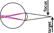

When a lens is focused just the right amount in front of a target, long wavelength light is focused sharply and short wavelength light is not. When a lens is focused just the right amount behind a target, short wavelength light is focused sharply.
The human eye introduces approximately 2 diopters of chromatic aberration across the visible range of the electromagnetic spectrum: 400 and 700 nm. Between the peak sensitivities of the long wavelength (L) and short (S) wavelength cone- 570 and 445 nm- chromatic aberration introduces 1 diopter of defocus. This is a huge amount of defocus. The defocus introduced by 1 diopter of chromatic aberration causes the same defocus blur as is caused by an object at 1 meter with your eye focused at 50 cm. Try it! Find an object 1 meter away, hold your finger at arm's length in front of the object, and focus on your finger. Note how blurry the object is. This blur is about equal the blur difference between the images captured by your L and S cones. We typically do not perceive color blurring because of our visual system's sophisticated processing of the retinal images. However, our visual system uses the signal to drive accommodation (biological auto-focusing) and estimate depth.
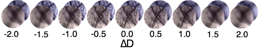
The images above show how chromatic aberration affects the blurriness of L and S cone images as a function of focus error. In broadband light, people focus preferentially on 570 nm light, the wavelength that L cones are maximally sensitive to. At 0 focus error, the L cone image (red tinted) is in best focus whereas the S cone image (blue tinted) is blurry (see ∆D=0.0). For positive focus errors (lens focused in front of target), the L cone image is sharper than the S cone image. For negative errors, the S cone image is sharper than the L cone image. The sharpness difference provide a signal to the sign of the focus error, and the overall sharpness of the images provides a signal to the magnitude of focus error.